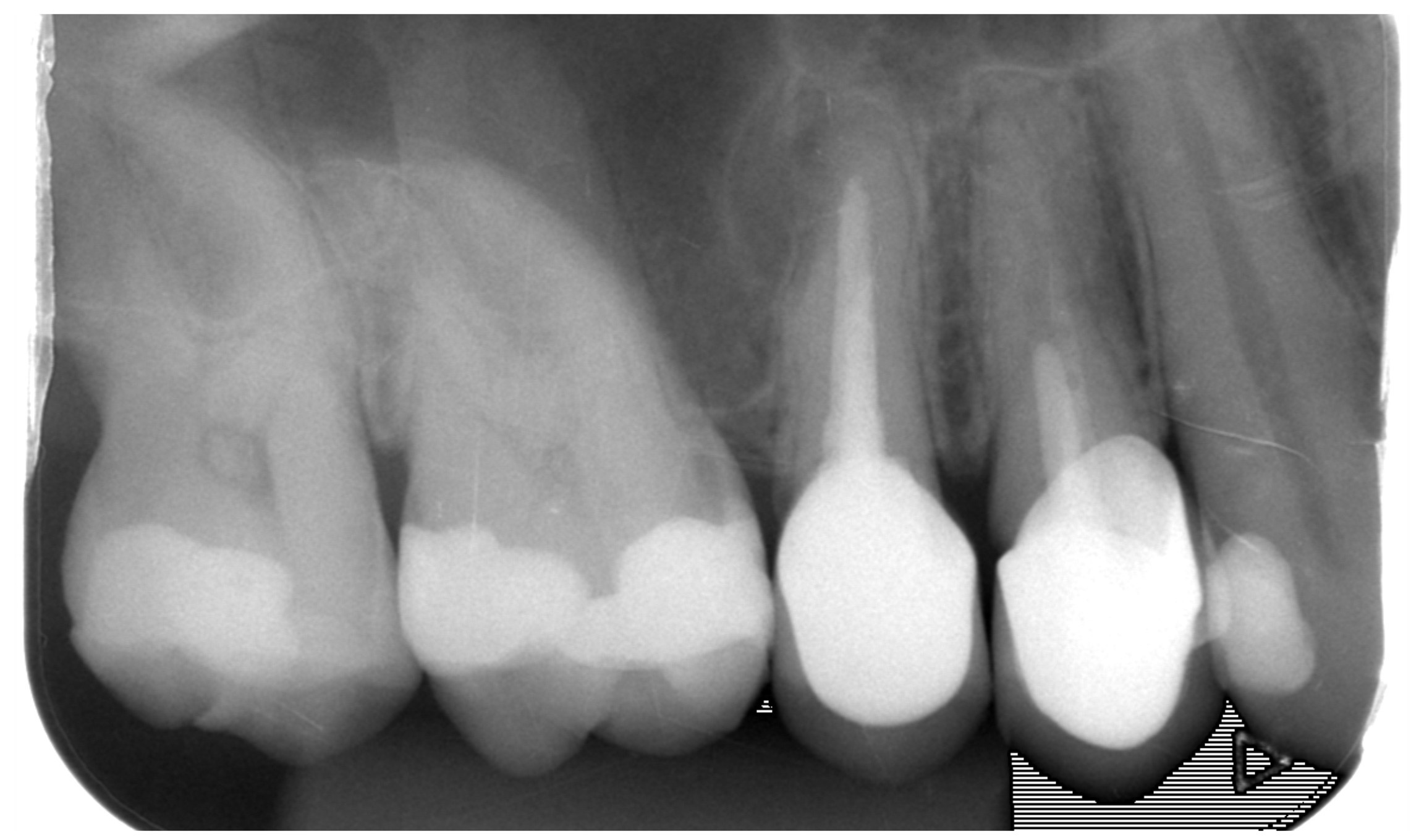
X-ray image before treatment: imprecise undercut crowns, periapical findings on both teeth ("pouches")

X-ray picture after removing the crowns and re-treatment of the root canals
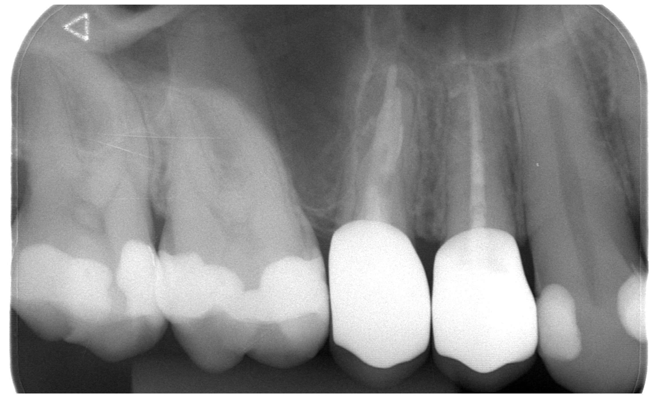
Control x-ray image – new crowns, healed periapical findings
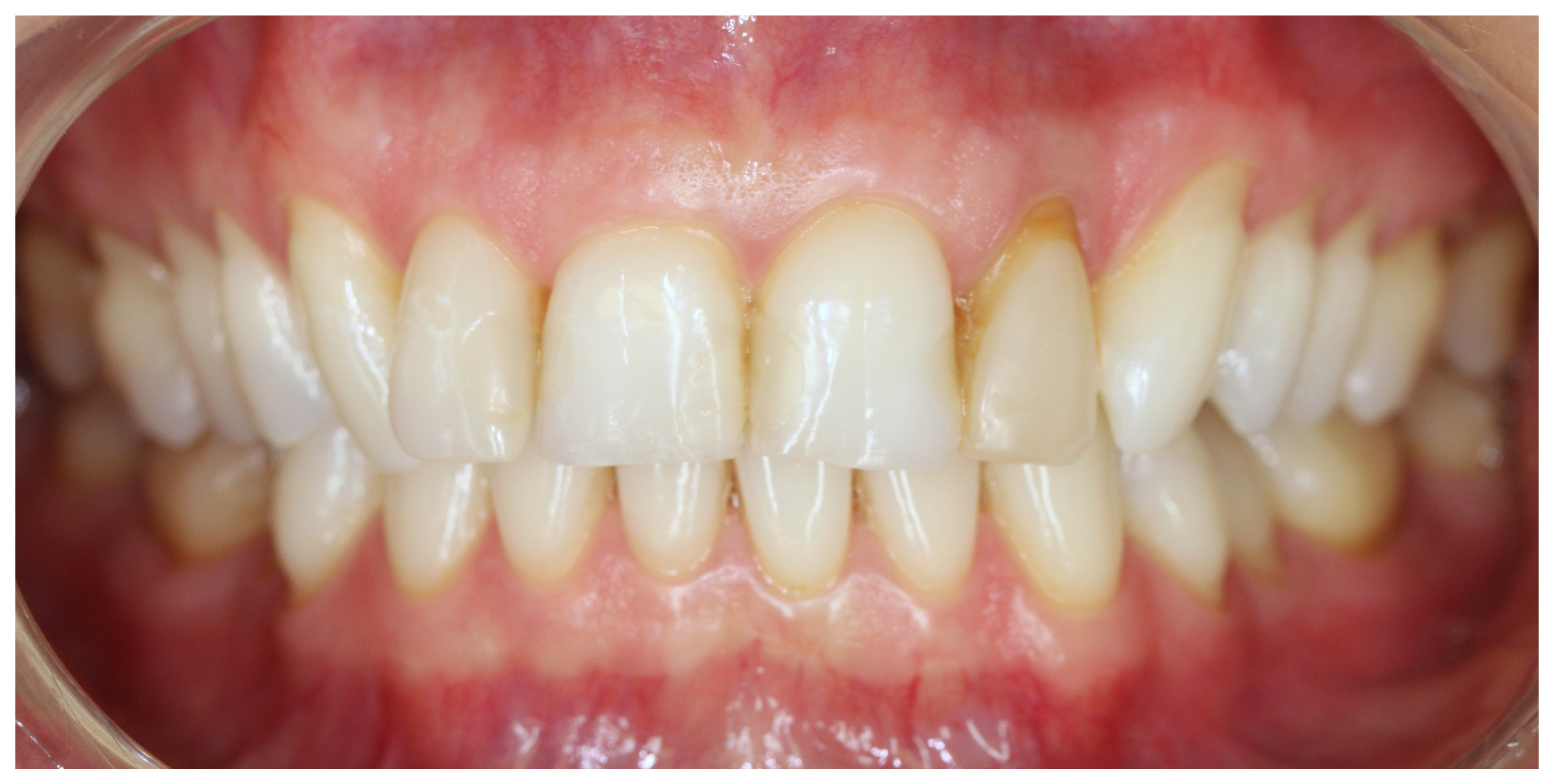
Initial condition before teeth whitening and before smile correction at the patient's request
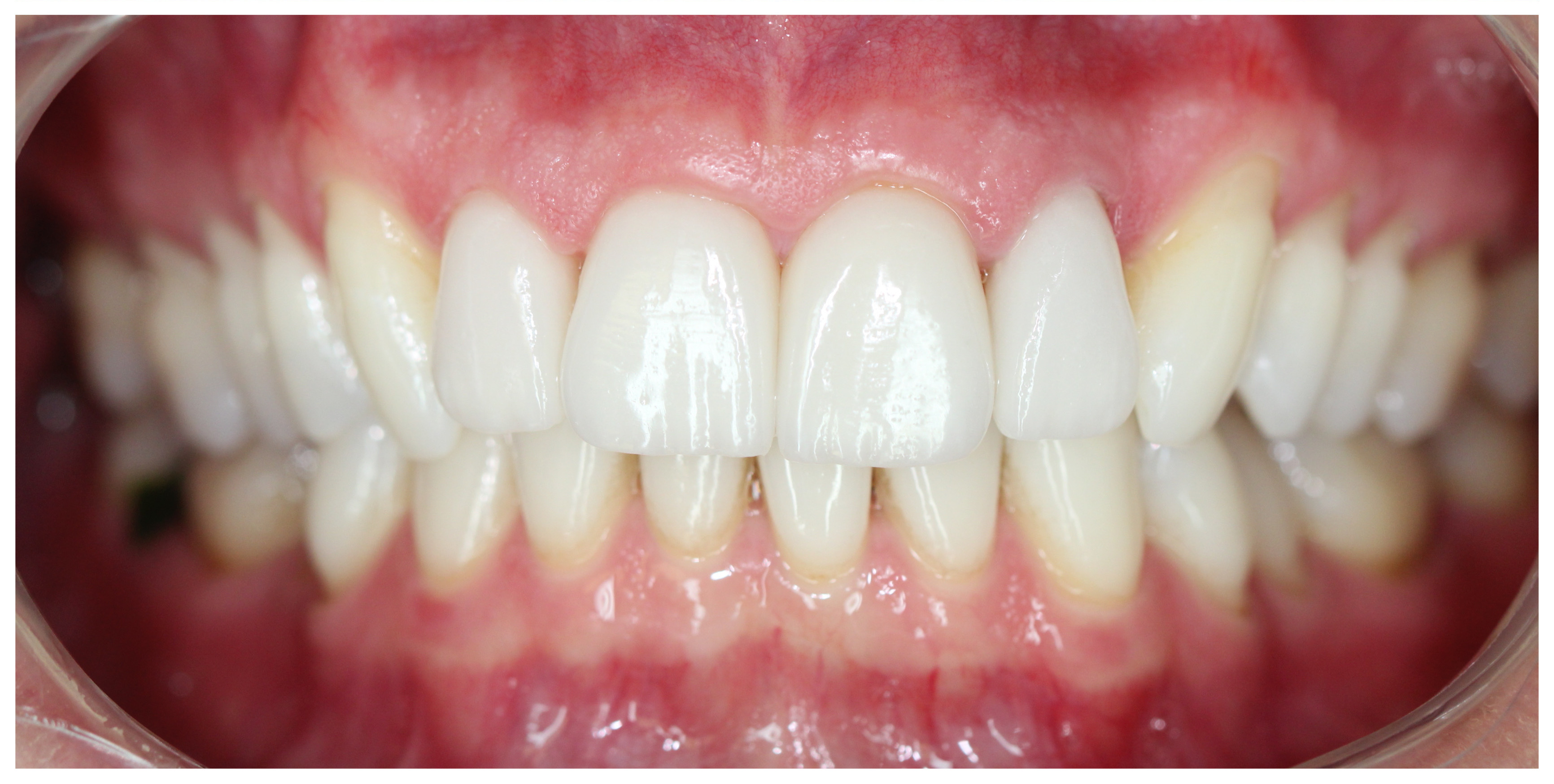
After teeth whitening and fabrication of three all-ceramic veneers and one crown on the upper incisors

OPG image at initial consultation - multiple cavities under old inaccurate fillings, periapical findings ("pouches"), poor endodontic treatment

OPG image taken three years after the end of rehabilitation - new endodontic treatment, new precise crowns and bridges, patient without problems

Undercut "white" filling on the six, new decay on the seven

New photocomposite fillings

OPG image at the initial consultation - new cavities and cavities under old fillings

Control OPG image two years after the end of the remediation

Excessively worn teeth due to orthodontic anomaly

Direct composite restorations after completion of orthodontic treatment
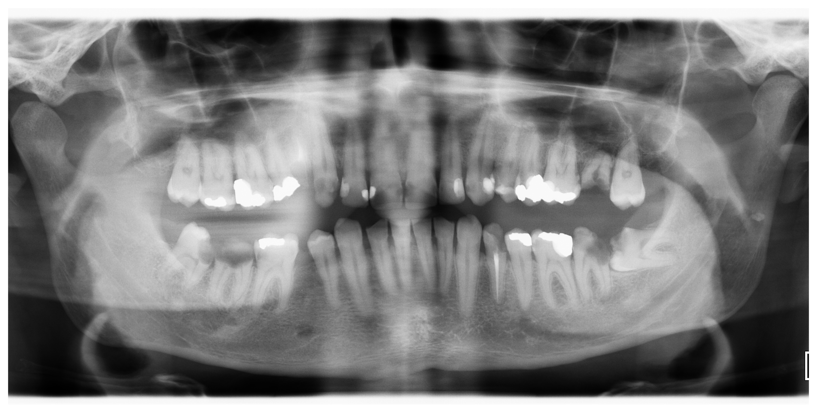
OPG image at the initial consultation – multiple caries and proven roots for extraction
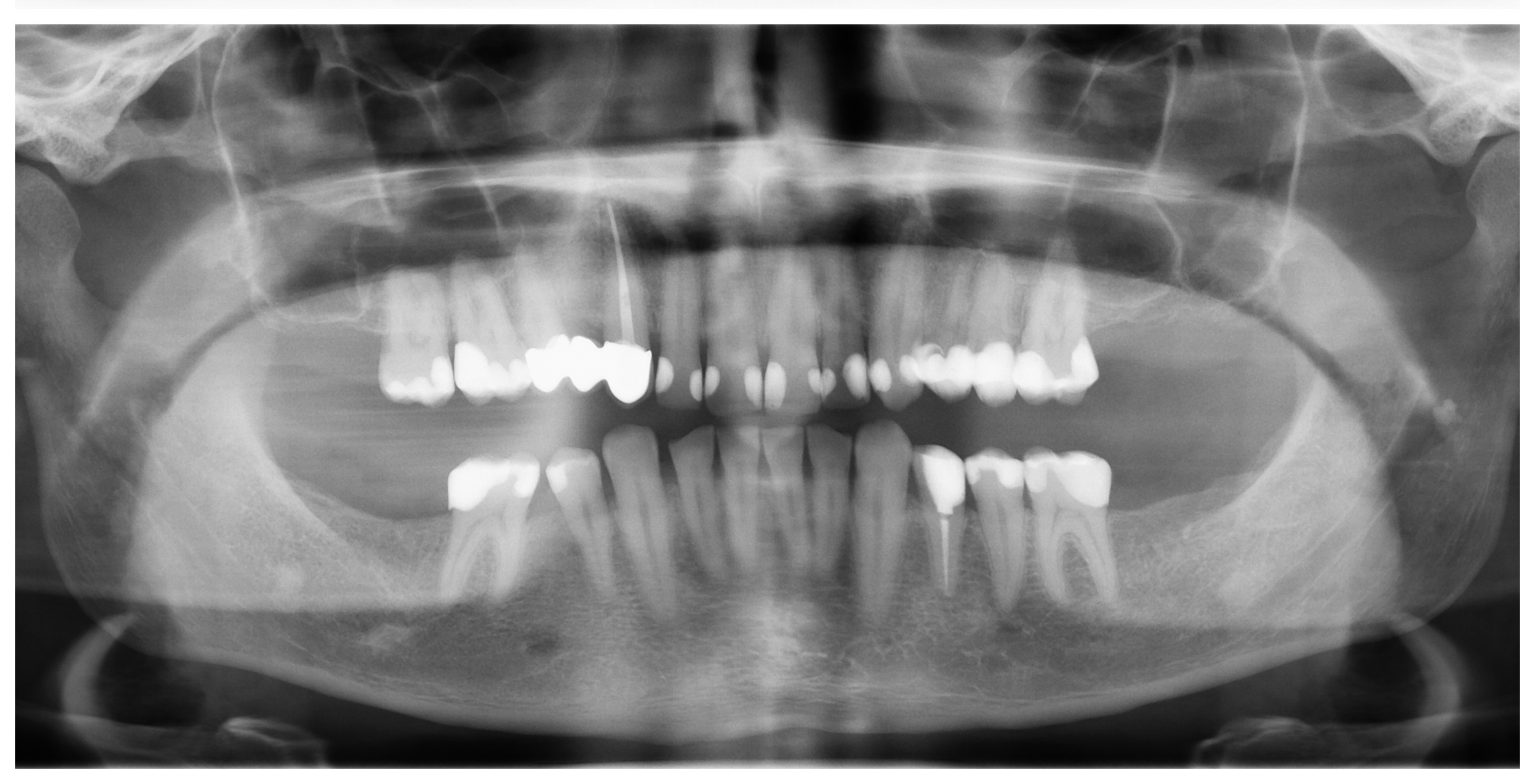
Control OPG image three years after the end of rehabilitation – long-term stable result, bone after extractions healed, defects repaired, gap after extracted upper quadrant replaced by a bridge

Provisional crowns on the upper twos, extensive composite completions of the ones

Definitive all-ceramic crowns on twos and ones

Primary caries on the upper six

Access to decay

Removed caries

Photocomposite filler

X-ray image before treatment of the upper five - insufficiently treated root canal, caries under the crown and inaccurate crown, undercut amalgam filling on the six and seven

X-ray picture after treatment - newly treated root canals, new precise crown on the fifth and photocomposite filling on the sixth and seventh

X-ray image after root canal treatment of the lower fifth with "pouch"

Control x-ray image after three months - "pouch" healed (+ new photocomposite filling on the six

X-ray image before treatment – caries on the five and seven and the six with caries and gangrene

X-ray picture after treatment - treated root canals on the sixth and fillings on the fifth, sixth and seventh